
1. SPoD-OnSPAN microscope
Super-resolution polarization demodulation/on-state polarization angle narrowing microscope
[ SPoD-OnSPAN microscope ]
A super-resolution fluorescence microscope to observe cells with very low phototoxicity. By our design of the microscope, observation method, and image reconstruction algorithm,
we have achieved super-resolution observation at a very low illumination power density so that we can perform real-time and long-term super-resolution observation of mammalian cells.
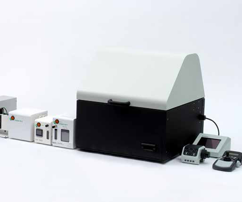
2. All-in-one bioluminescence microscope [PDF]
[ All-in-one bioluminescence microscope ]
A high-magnification microscope to observe bioluminescence in highly-dark condition.
The construction is underway.
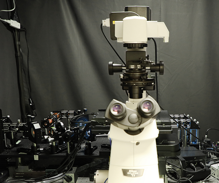
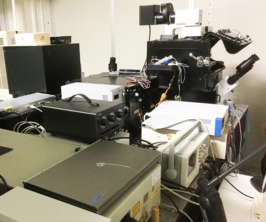
3. Single-molecule fluorescence microscope
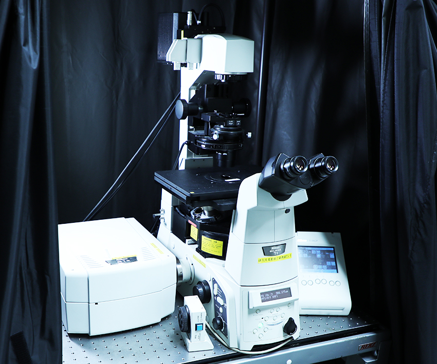
[ Single-molecule fluorescence microscope ]
A total internal reflection fluorescence (TIRF) microscope for single-molecule imaging. The in-house built illumination optical system includes 405, 488, 561, and 640 nm lasers, and is combined with a Nikon Ti inverted microscope. On the microscope,
we also perform super-resolution imaging by single-molecule localization microscopy (SMLM).
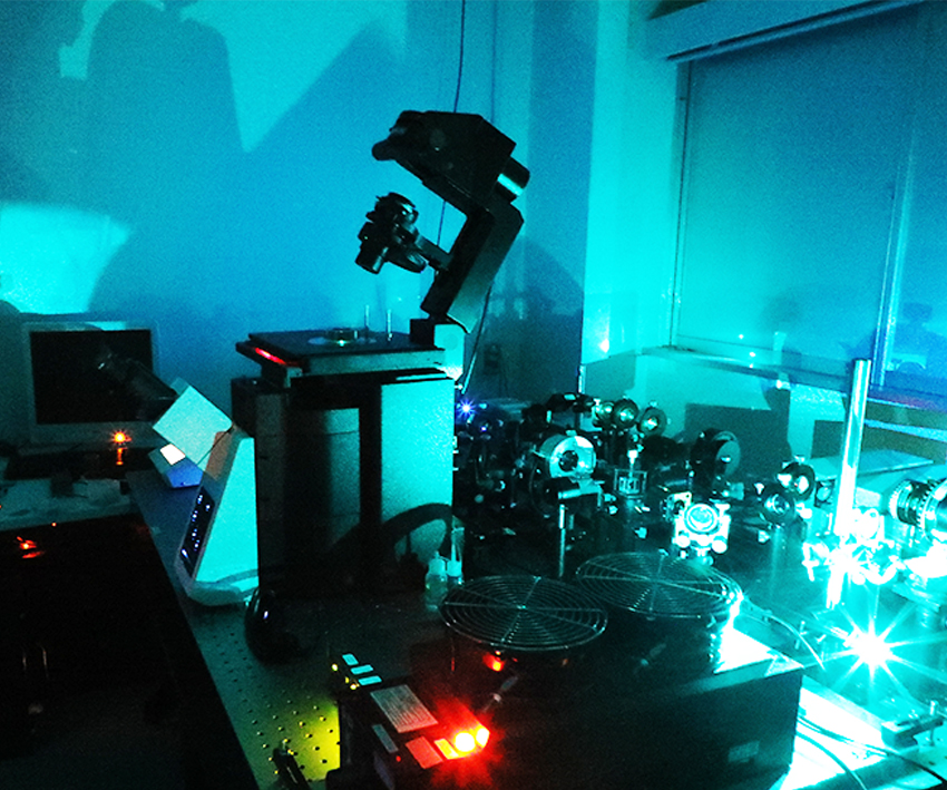
4. Cavity-reflection-enhanced absorption microscopy (CREAM)
[ Cavity-reflection-enhanced absorption microscopy (CREAM) ]
A microscope to map absorbance at super-enhanced sensitivity. A light beam is allowed to pass through a specimen many times in our uniquely-developed optical cavity to enhance the contrast of very low absorbance.
Being detected by a camera, the spatial distribution of the enhanced light absorption is visualized.
5. Incoherent holography microscope
[ Incoherent holography microscope ]
By incoherent digital holography, a few frames of interference patterns from the fluorescence or chemiluminescence of cells or tissues are taken to reconstruct a 3D image.
The construction of this microscope is now underway.
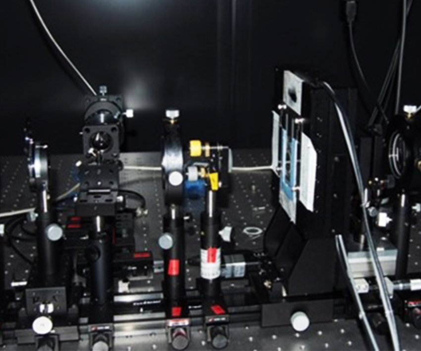
6. Scanning two-photon excitation fluorescence microscope
[ Scanning two-photon excitation fluorescence microscope ]
A fluorescence microscope to observe cells and tissues at extended depth. By the technique of two-photon excitation with near infrared laser beam,
fluorescence in a thick specimen can be observed. This microscope is also used to take a 3D image.
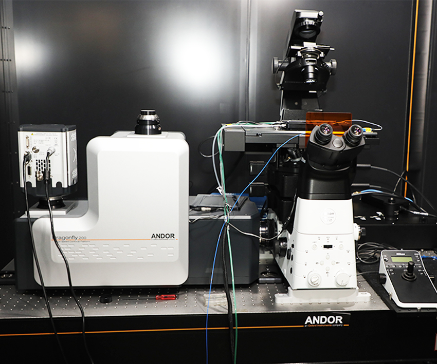
7. Next generation multi-beam high-speed confocal fluorescence microscope
Nikon/Andor Dragonfly
[ Next generation multi-beam high-speed confocal fluorescence microscope ]
A microscope system composed of a Nikon Ti2E inverted microscope with a super-wide FOV and an Andor Dragonfly high-speed confocal module.
Highly-sensitive real-time 3D observation and SRRF super-resolution imaging are performed with an EMCCD camera and high-speed scanning of various wavelengths of laser beams.
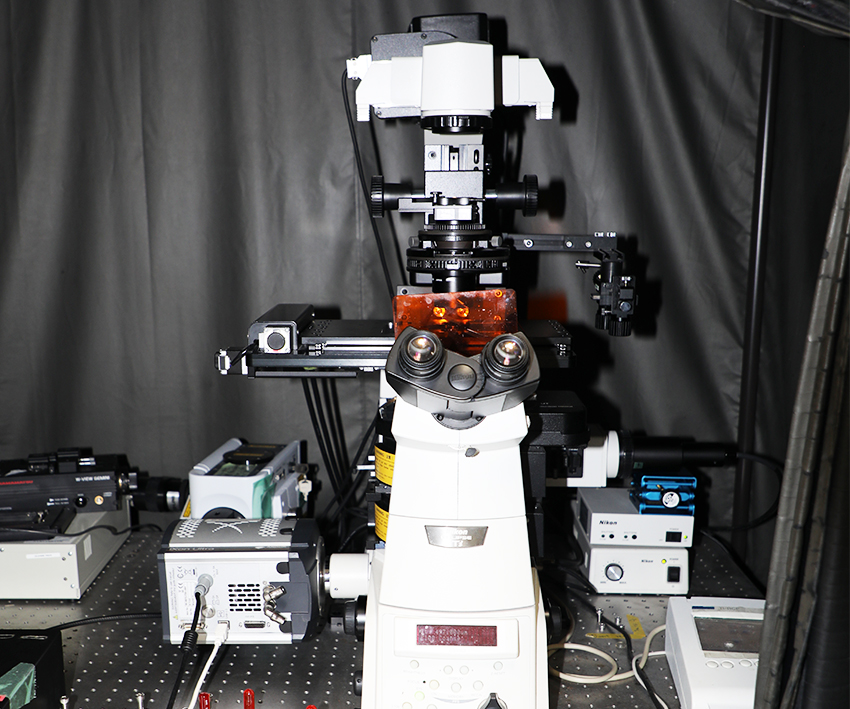
8. Multi-focus multi-color confocal microscope
Nikon/OPTO-LINE
[ Multi-focus multi-color confocal microscope ]
A multiple-purpose confocal fluorescence microscope.
This is composed of a Nikon Ti inverted microscope and an Optoline MESSIA confocal module with a Nipkow disk and LEDs.
9. High-speed spectral confocal microscope
[ High-speed spectral confocal microscope ]
A laser-scanning confocal microscope with spectroscopy capability.
This includes a Nikon Ti inverted microscope and an A1R confocal module. High-speed confocal observation and light stimulation with 405 nm beam can be simultaneously performed.
10. Spectral confocal microscope
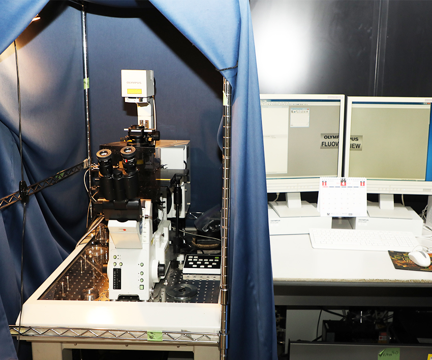
[ Spectral confocal microscope ]
An Olympus FLUOVIEW FV1000 laser-scanning confocal microscope. This microscope performs not only confocal observation but also simultaneously light stimulation with the SIM scanner unit.
Furthermore, this is equipped with a fluorescence spectrophotometer with a 2-nm resolution.
11. A Keyence all-in-one fluorescence microscope BZ-X700
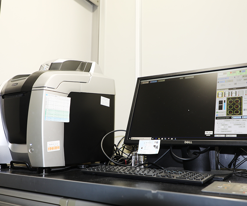
[ A Keyence all-in-one fluorescence microscope BZ-X700 ]
This highly-functional and highly-user-friendly microscope performs fluorescence,
bright-field, and phase-difference contrast observation. On this microscope, an auto-focus mechanism, a motorized stage, and the automation of observation sequences are also available.
12. Bioluminescence microscope
OLYMPUS LV200
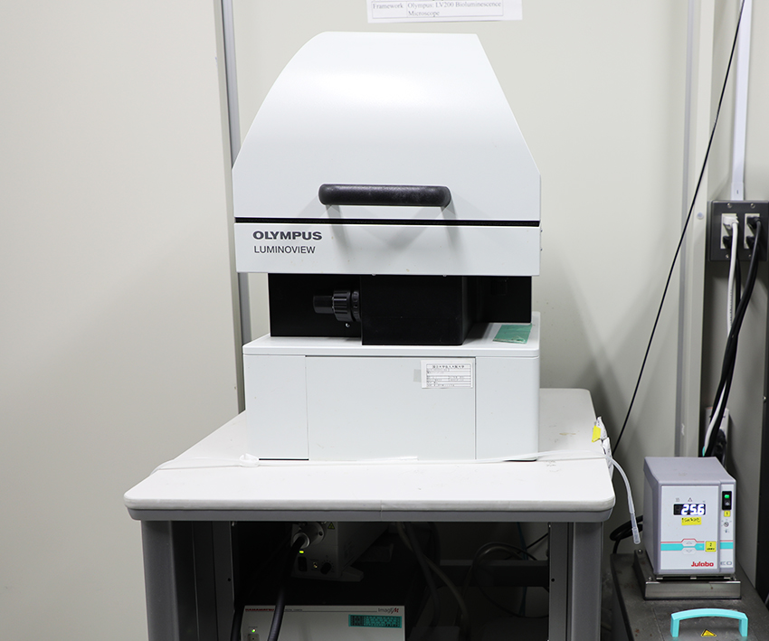
[ Bioluminescence microscope ]
An Olympus LV200 microscope for bioluminescence observation. This is equipped with a metal cover, which enables dark-conditioned observation free of the room lighting.
Furthermore, the optimized optical system and peripherals supports highly-sensitive observation of cells emitting bioluminescence.
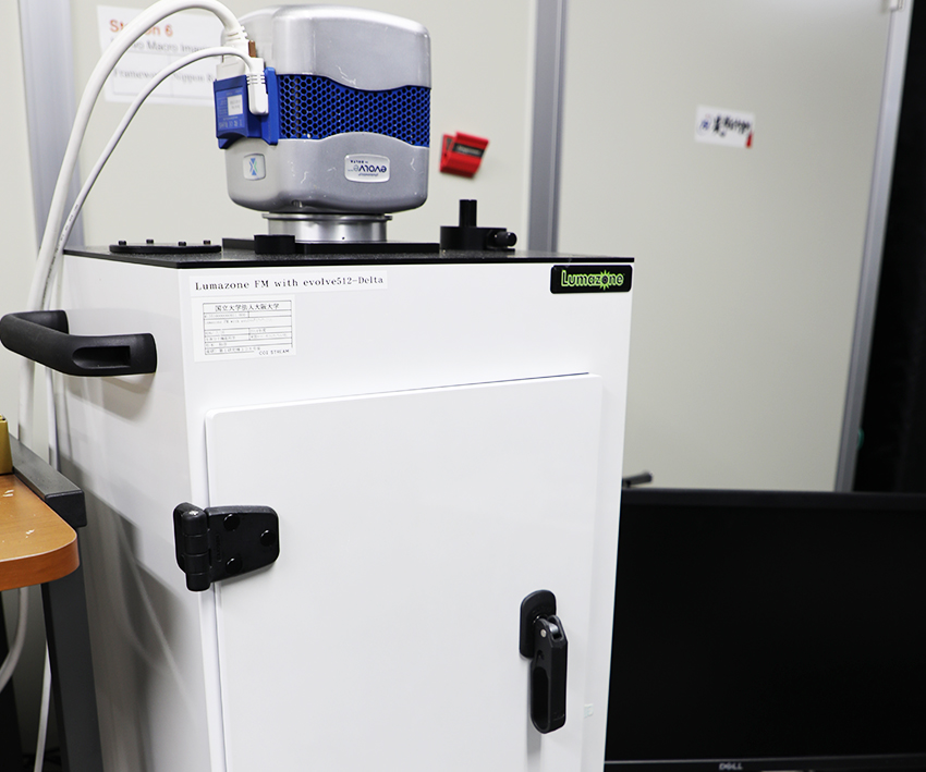
13. In vivo bioluminescence imaging system
[ In vivo bioluminescence imaging system ]
Lumazone CMS, an in vivo bioluminescence imaging system by Nippon Roper. Animals and plants are observed with minimal stress and disturbance in the highly-dark condition.
With an EMCCD, bioluminescence can be detected at high sensitivity.
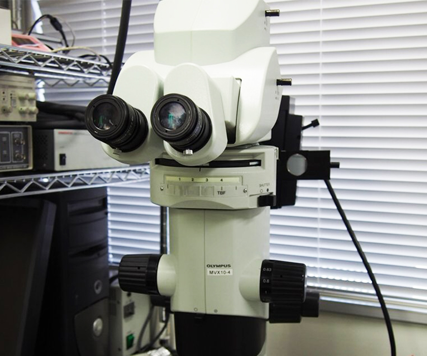
14. Macro zoom microscope
MVX10 ( Olympus )
[ Macro zoom microscope ]
An Olympus MVX10 macro zoom microscope.
This high-numerical-aperture microscope performs fluorescence and bright-field observation from cells at a high magnification to animals and plants at a low magnification.
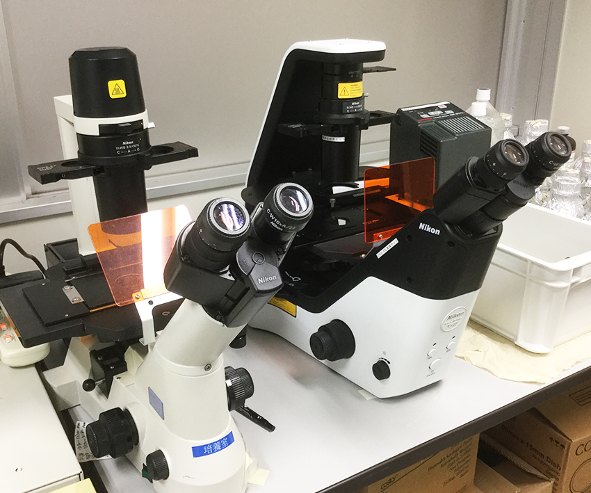
15. Fluorescence and phase difference contrast microscopes
[ Fluorescence and phase difference contrast microscopes ]
These are Nikon TS2 and TS100 inverted microscopes. Being next to a CO2 incubator, they are used to perform fluorescence, phase-difference contrast,
and bright-field observation for not just cultured cells expressing fluorescent proteins but also unlabeled cells.
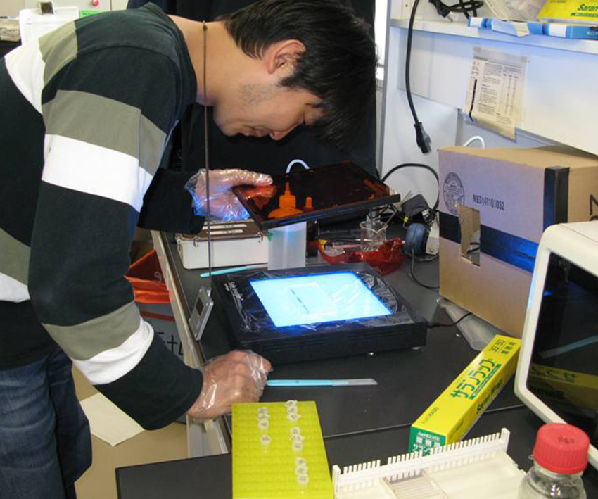
1. Invitrogen Safe Imager
青色光トランスイルミネーター
Invitrogen Safe Imager
ゲルからのDNAの切り出しに使用.
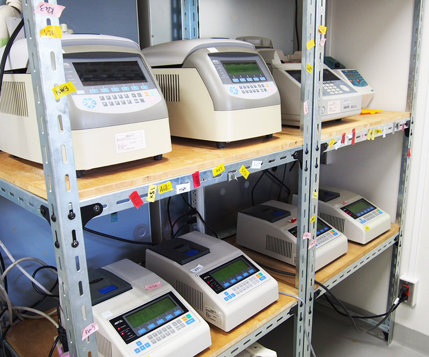
2. PCR装置
PCR装置
DNAの増幅に使用.
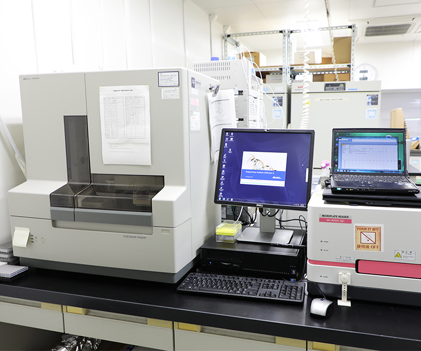
3. DNA シーケンサー
ABI PRISM 310 Genetic Analyzer
DNA シーケンサー
DNA配列の決定に使用.
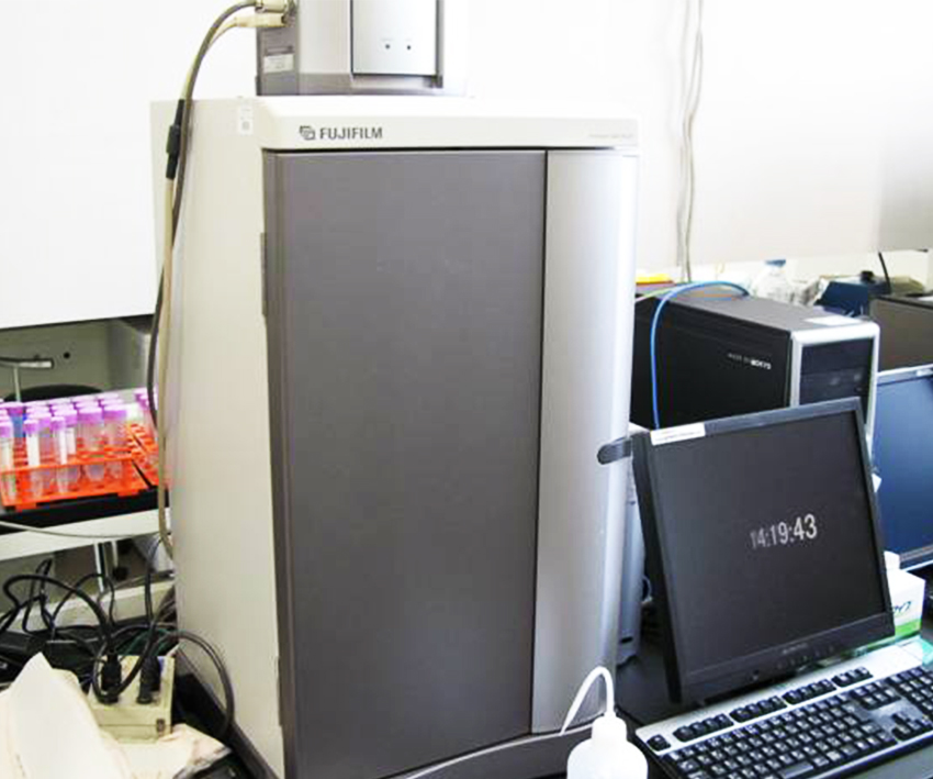
4. ルミノ・イメージアナライザー
FUJIFILM LAS-1000plus
ルミノ・イメージアナライザー
蛍光, 発光サンプルのデジタル画像取得に使用.
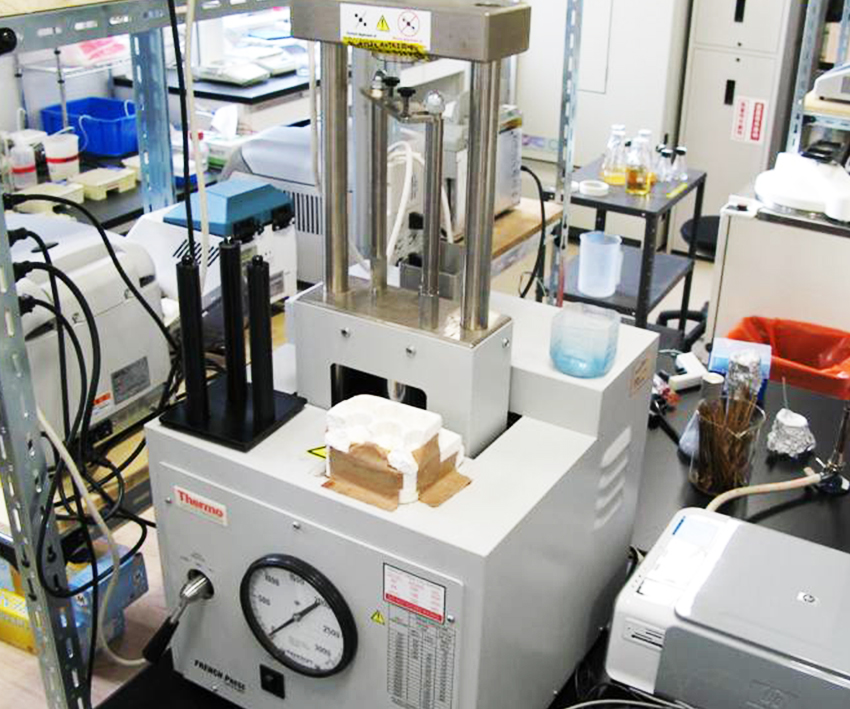
5. フレンチプレス
フレンチプレス
細胞・菌体の破砕に使用.
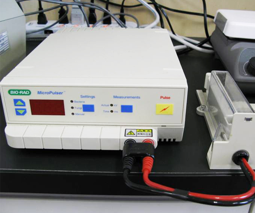
6. エレクトロポレーター
Bio-rad MicroPulser
エレクトロポレーター
電気パルスでDNAを細胞に導入するのに使用.
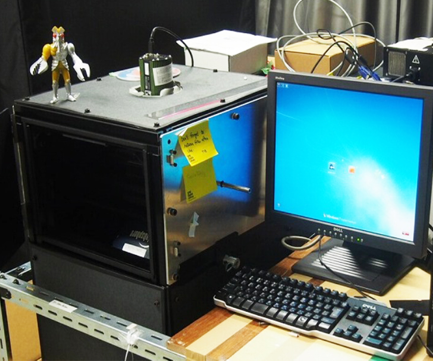
7. バルタン
独自開発 自作蛍光スクリーニング装置
バルタン
蛍光タンパク質のスクリーニングの際に使用. 積分球を利用して均一な励起光を照射することで, 容易に蛍光タンパク質のスクリーニングを行うことが可能.
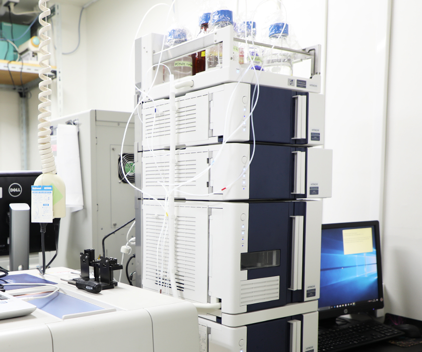
8. 高速液体クロマトグラフィーシステム
HITACHI 高速液体クロマトグラフChromaster
[ 高速液体クロマトグラフィーシステム ]
日立ハイテクサイエンス製高速液体クロマトグラフChromaster. 主にゲル濾過,分配,イオン交換の分離モードで分析を行っている.
PEEKおよびテフロンの接液部仕様によってタンパク質の生理活性に対する影響を最小化している. フォトダイオードアレー検出器により分析中にリアルタイムで吸光スペクトルをモニターできる.
1. 分光蛍光光度計
JASCO FP-8300 ストップトフローシステム:SFS
[ 分光蛍光光度計 ]
励起・蛍光スペクトル測定に使用. また高感度測定と広いダイナミックレンジを有しており, 波長走査速度による高速3Dスペクトル測定などにも使用.
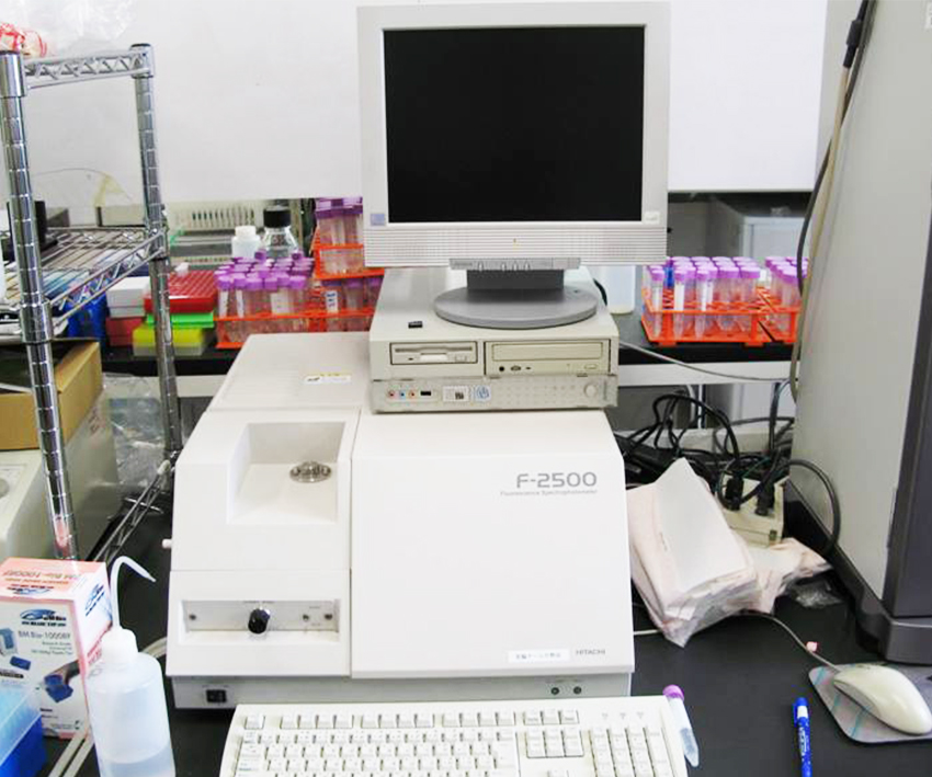
2. 蛍光分光光度計
HITACHI F-2500
[ 蛍光分光光度計 ]
励起・蛍光・発光スペクトル測定に使用.
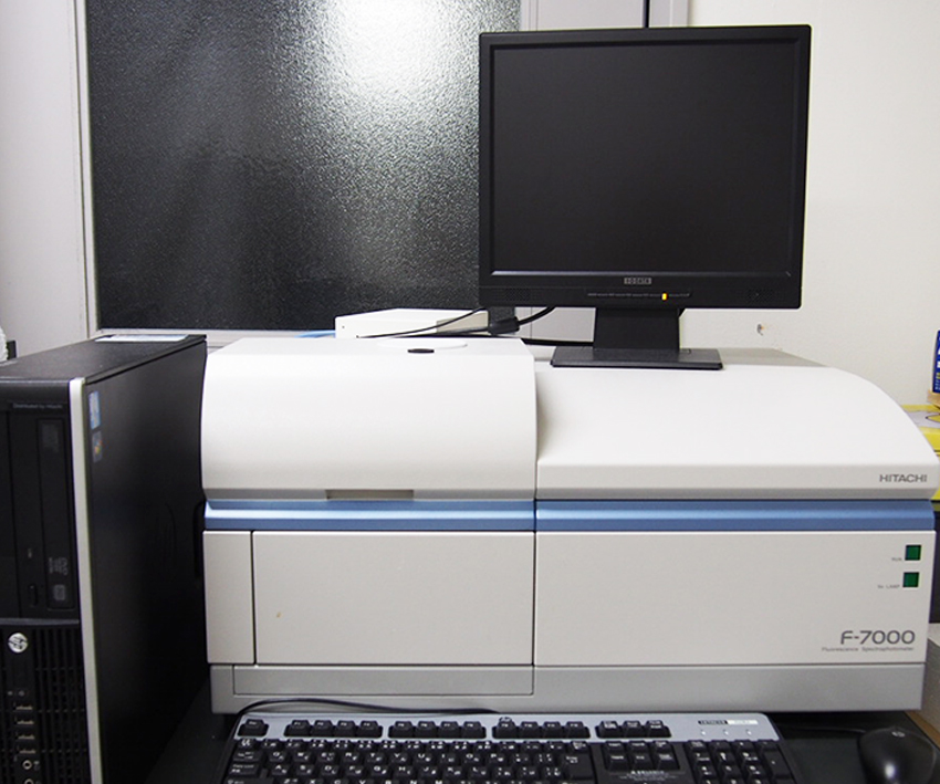
3. 蛍光分光光度計
HITACHI F-7000
[ 蛍光分光光度計 ]
励起・蛍光・発光スペクトル測定に使用.
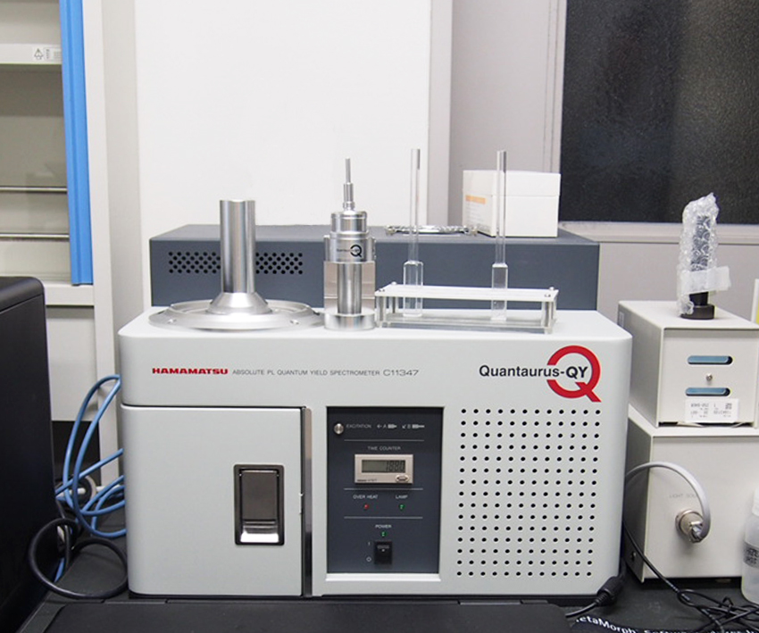
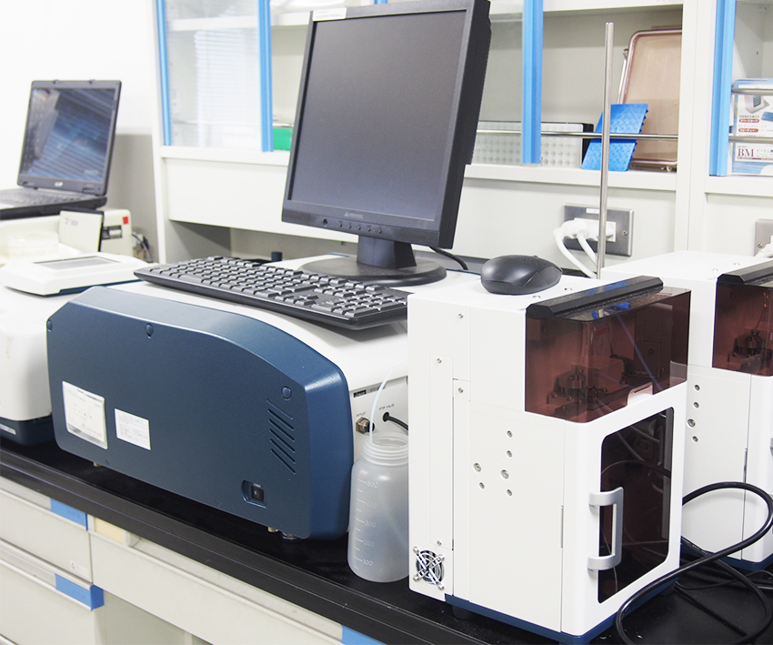
4. 絶対蛍光量子収率測定装置
[ 絶対蛍光量子収率測定装置 ]
フォトルミネッセンス法によって、発光量子収率の絶対値を瞬時に測定できる装置.
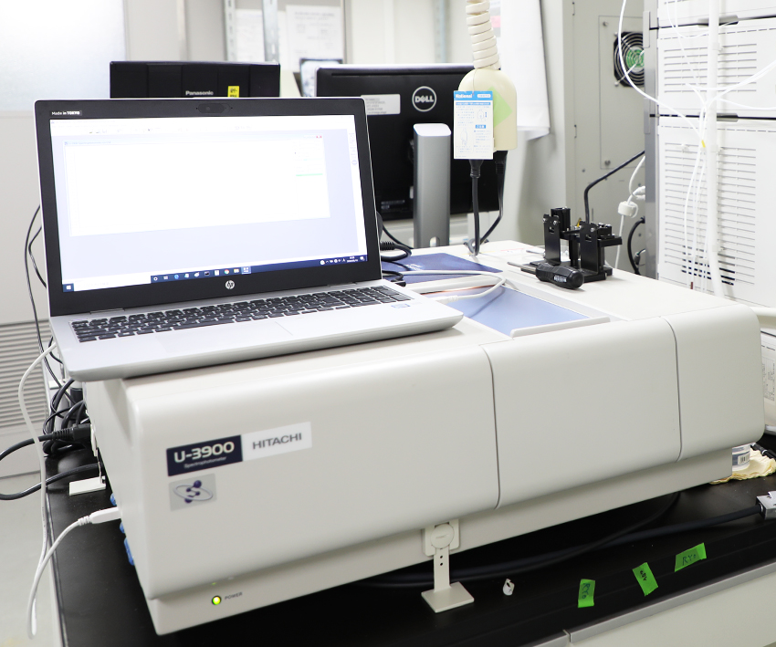
5. 吸光分光光度計
HITACHI U-3900
[ 吸光分光光度計 ]
日立ハイテクサイエンス製紫外可視分光光度計U-3900. タンパク質,DNA,生理物質,光学フィルター等の光吸収の分光特性を高分解能高感度で測定する.
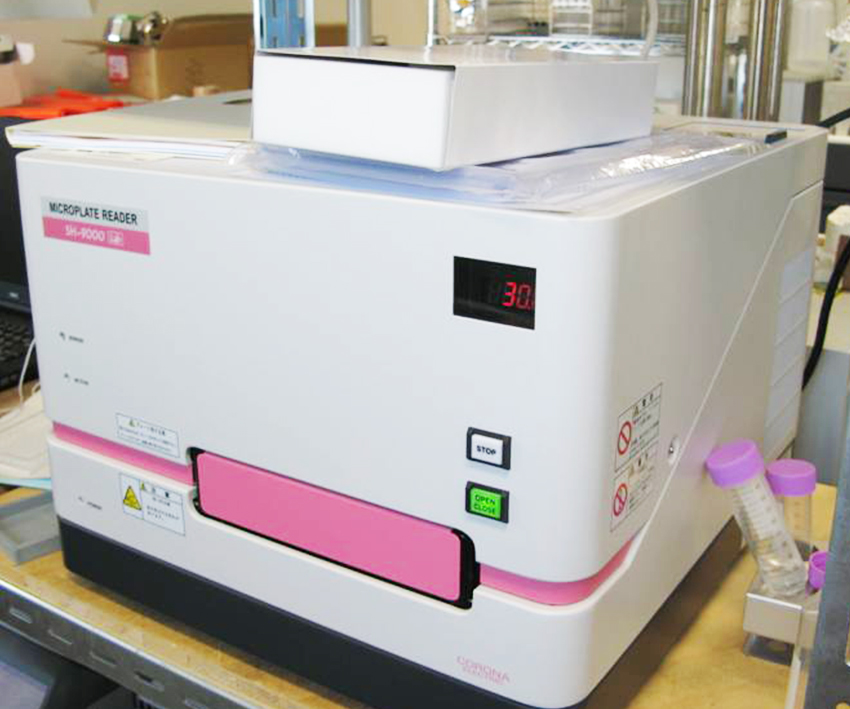
6. マイクロプレートリーダー CORONA
GRATING MICROPLATE READER SH-9000
[ マイクロプレートリーダー CORONA ]
ハイスループットに吸光・蛍光・発光・時間分解蛍光測定を行う.
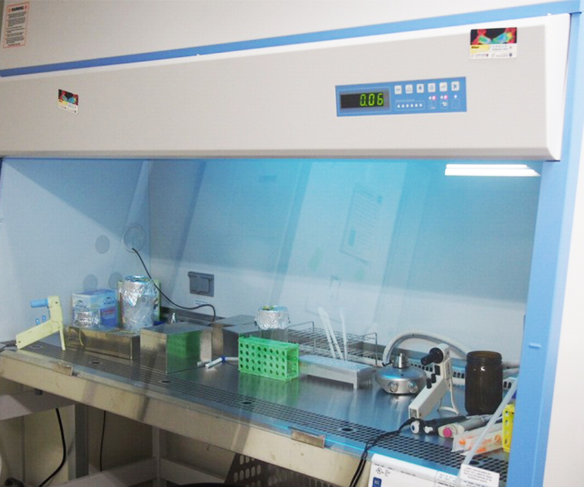
1. 安全キャビネット
[ 安全キャビネット ]
バイオハザードを封じ込め, 安全な作業環境を実現する設備.
安全キャビネットは内部の試料が外部へ漏れるのを防止のために内部を低圧に保ち, またフィルターを通して清浄化した空気を排出.
2. 粘菌培養
[ 粘菌培養 ]
実験生物の粘菌を培養.